
X-Ray Digital Radiography and Computed Tomography
for Cultural Heritage
Franco Casali
Department of Physics, University of Bologna
Tel. +39 051 2095131 – 253274
E-mail address: casali@bo.infn.it
X-ray detection systems for high resolution Digital Radiography (DR) and Computed Tomography (CT) have been developed at the Physics Department of the University of Bologna. The research target is the development of systems to be applied in cultural heritage conservation and industrial radiology.
In the field of cultural heritage, different kind of objects (ancient necklaces, paintings, bronze or marble statues) have to be inspected in order to acquire significant information as the method used to assemble, the manufacturing techniques or the presence of defects. These features could be very useful, for example, for dating works of art or determining appropriate maintenance and restoration procedures. Among the advanced methods available, 3D CT can be successfully used for the investigation of ancient works of art because it preserves their integrity and provides images of inner parts, otherwise not visible.
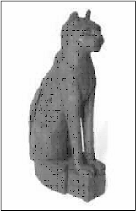
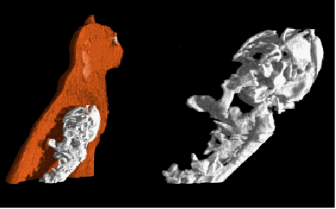
Several high-resolution CT systems to investigate objects of different sizes (from micro to macro) have been developed. For example, we have carried out the micro CT reconstruction of Roman human tooth with carie (found in the “Isola Sacra” necropolis); as well as the cone beam CT analysis on an Egyptian cat-shaped coffin exhibiting the inner mummy; until to the CT study of an ancient large globe (2 m of diameter). This globe was created by a Dominican monk, Egnazio Danti, around 1567 and is located in Palazzo Vecchio, at Florence. The 3D CT reconstruction of the globe clearly shows the entire inner structure, consisting of a central pole, 8 bars as 2 tetrahedrons and 30 meridians. All the inner structure is made of iron, with a total weight of about 350 kg, estimated from the segmented 3D reconstruction. The very high resolution reached investigating small objects is an important result other than tomography on a big object, like the globe, is an absolute innovation. A 3D CT investigation is being in project to determine how much deterioration has occurred on the ankles of David, the towering marble figure sculpted by Michelangelo, a very exciting purpose that we will achieve in collaboration with Lawrence Livermore National Laboratory.
A new linear array detector for high resolutions and low dose digital radiography for painting was realized. This new instrument is able to acquire radiological image with an amount of dose one hundred times reduced. The system was tested on a benchmark panel with a number of classified original pigments provided by the "Opificio delle Pietre Dure" Institute in Florence. The resolution and the image contrast reached by the scanning system were superior to that of the common film systems used at the Institute.
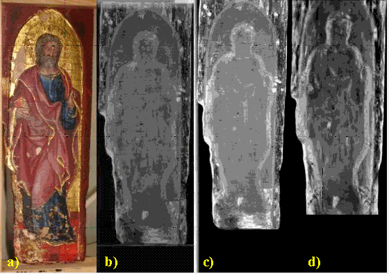
Moreover, in collaboration with the National Gallery of Bologna and the "Opificio dele Pietre Dure" restoration center in Florence, was performed an X-ray investigation of the inner structure of two small (20 cm 7 cm) painted "tablets", made of wood, and recognized as an artwork of Gentile da Fabriano, an important painter of the XIII century. Different techniques were used: conventional film radiography, digital radiography and computed tomography, the latter two with innovative equipment of the Department of Physics in Bologna. An important result was achieved with the intensified fiber optics scanner. A quality close to that of a conventional film was obtained with estimated two orders of magnitude less dose. The x-ray scanner developed at Department of Physics demonstrated to be a very useful and vanguard tool for direct digital radiography of paintings.
We also have been performed experiments with multispectral imaging equipments. These techniques are powerful to investigate ancient book in order to support the study of library materials, the mechanisms involved in deterioration, to discover hidden information and to assist book conservation and restoration.


